Multiplex Antibody Detection
What Is Multiplex Antibody Detection?
Multiplex antibody detection refers to the simultaneous detection of two or more targets using antibodies and an appropriately designed analytical system.
In a conventional single-target assay, one antibody-based detection system is used to investigate a specific antigen. In a multiplex assay, several targets are examined together, allowing researchers to compare their presence, distribution, or abundance within the same sample.
For example, researchers studying inflammation may want to investigate several cytokines or immune cell markers at once. A multiplex assay can help characterize these targets within a shared experimental framework, depending on the technology and its validated measurement range.
Multiplex detection can be performed using different techniques, including multiplex immunofluorescence, flow cytometry, bead-based immunoassays, and multiplex ELISA platforms. Each method has specific requirements for antibody compatibility, signal separation, sensitivity, and data interpretation.

How Does Multiplex Antibody Detection Work?
The basic principle involves using antibodies that recognize different targets and assigning each target a distinguishable detection signal or measurement channel.
The exact workflow depends on the assay format, but most multiplex experiments involve several essential stages.
1. Target Selection
Researchers first identify the proteins, biomarkers, or cellular structures they want to investigate. Targets should be relevant to the research question and detectable using the selected analytical method.
2. Antibody Selection
Each target requires an antibody with suitable specificity and validated performance. Researchers may select monoclonal or polyclonal antibodies according to the experimental requirements.
The host species of each primary antibody is particularly important in indirect detection systems because secondary antibodies must distinguish the primary reagents without producing unwanted signals.
3. Detection Label Selection
Different targets can be associated with distinct fluorescent dyes, enzymes, or other detectable labels. The selected signals must be distinguishable using the available instrumentation.
For fluorescence-based experiments, researchers consider excitation and emission spectra, channel overlap, sample autofluorescence, and the sensitivity of the detection equipment.
4. Signal Acquisition
The sample is analyzed using an appropriate instrument, such as a fluorescence microscope, flow cytometer, or microplate reader. The instrument records the signals associated with the different targets.
5. Data Analysis
Researchers evaluate the individual signals, assess background levels, and compare results with suitable controls. Depending on the assay, analysis may involve fluorescence intensity, target concentration, cellular distribution, or the proportion of cells expressing particular markers.
The Importance of Antibody Host Species
Antibody host species refers to the animal in which an antibody was generated, such as a rabbit, mouse, goat, rat, or chicken. This characteristic is particularly important when designing multiplex experiments that rely on secondary antibody detection.
For example, a rabbit primary antibody can be detected using a compatible anti-rabbit secondary antibody. A mouse primary antibody can be detected separately using an anti-mouse secondary antibody, provided that the reagents and experimental conditions are suitable.
Using primary antibodies from different host species can simplify the separation of signals in indirect detection systems. However, species separation alone does not guarantee specificity. Secondary antibody cross-reactivity, endogenous immunoglobulins, sample composition, and the properties of the detection system must also be considered.
When multiple primary antibodies originate from the same species, conventional secondary antibodies may bind to more than one primary antibody, making it difficult to assign signals to individual targets.
Possible solutions include using directly conjugated primary antibodies, validated sequential staining protocols, specialized detection systems, or other methods designed to distinguish the targets.


Monoclonal and Polyclonal Antibodies in Multiplex Assays
The choice between monoclonal and polyclonal antibodies can influence the specificity, sensitivity, and reproducibility of multiplex detection.
Monoclonal Antibodies
Monoclonal antibodies are derived from a defined antibody-producing clone or sequence and generally recognize a specific epitope. Their defined recognition properties can be useful when researchers need to distinguish several targets within the same experiment.
In multiplex assays, monoclonal antibodies may help support consistent target recognition, provided that each reagent has been validated for the intended application.
Polyclonal Antibodies
Polyclonal antibody preparations contain multiple antibody populations that recognize different epitopes on the same antigen. They may provide strong signals in certain assays, but their broader recognition profile can introduce additional specificity considerations.
When polyclonal antibodies are included in multiplex experiments, researchers should evaluate their specificity, potential cross-reactivity, and compatibility with the other reagents.
Neither format is universally superior. The best choice depends on the targets, assay design, sample type, and available validation data.
Major Techniques Used in Multiplex Antibody Detection
Multiplex Immunofluorescence
Multiplex immunofluorescence uses fluorescently labeled antibodies to visualize several targets within cells or tissues. Different fluorescent signals can reveal the localization of multiple proteins in the same sample.
This technique is valuable for studying tissue organization, immune cell populations, protein distribution, and interactions between biological structures.
Successful experiments require appropriate fluorophore selection, control of spectral overlap, and validated staining protocols.
Multiparameter Flow Cytometry
Flow cytometry analyzes individual cells according to their physical properties and fluorescent signals. Multiparameter experiments use several labeled antibodies to identify multiple cellular markers simultaneously.
Researchers can use this approach to characterize immune cell populations, compare marker expression, and investigate cellular heterogeneity.
Important considerations include fluorophore compatibility, compensation, antibody titration, instrument configuration, and appropriate controls.
Multiplex ELISA
Multiplex ELISA platforms enable the measurement of multiple analytes in a sample. Depending on the technology, targets may be distinguished through separate capture regions, spatially separated assay locations, or other signal-identification systems.
These assays are useful for measuring groups of proteins, including cytokines, chemokines, and other biomarkers.
The antibodies must recognize compatible epitopes, and each analyte should be assessed for sensitivity, specificity, interference, and performance within the validated measurement range.
Bead-Based Multiplex Immunoassays
Bead-based platforms use distinguishable particle populations associated with specific capture reagents. Each bead population corresponds to a particular analyte, allowing several targets to be measured in a shared sample.
These methods can support the analysis of multiple biomarkers while conserving sample volume. Their performance depends on the assay platform, reagent compatibility, calibration, and data analysis procedures.


Advantages of Multiplex Antibody Detection
Multiplex detection offers several potential benefits when the assay is appropriately designed and validated.
Efficient analysis: Multiple targets can be investigated within a shared experimental workflow.
Reduced sample consumption: Some multiplex platforms require less sample than performing separate single-target assays.
Comprehensive biological information: Researchers can evaluate several related markers together.
Improved experimental comparisons: Measuring multiple targets under similar conditions can facilitate the investigation of relationships between biological signals.
Time efficiency: Combining measurements may reduce the time required compared with running multiple separate assays.
These advantages depend on the platform, sample requirements, assay complexity, and performance of the individual detection systems.
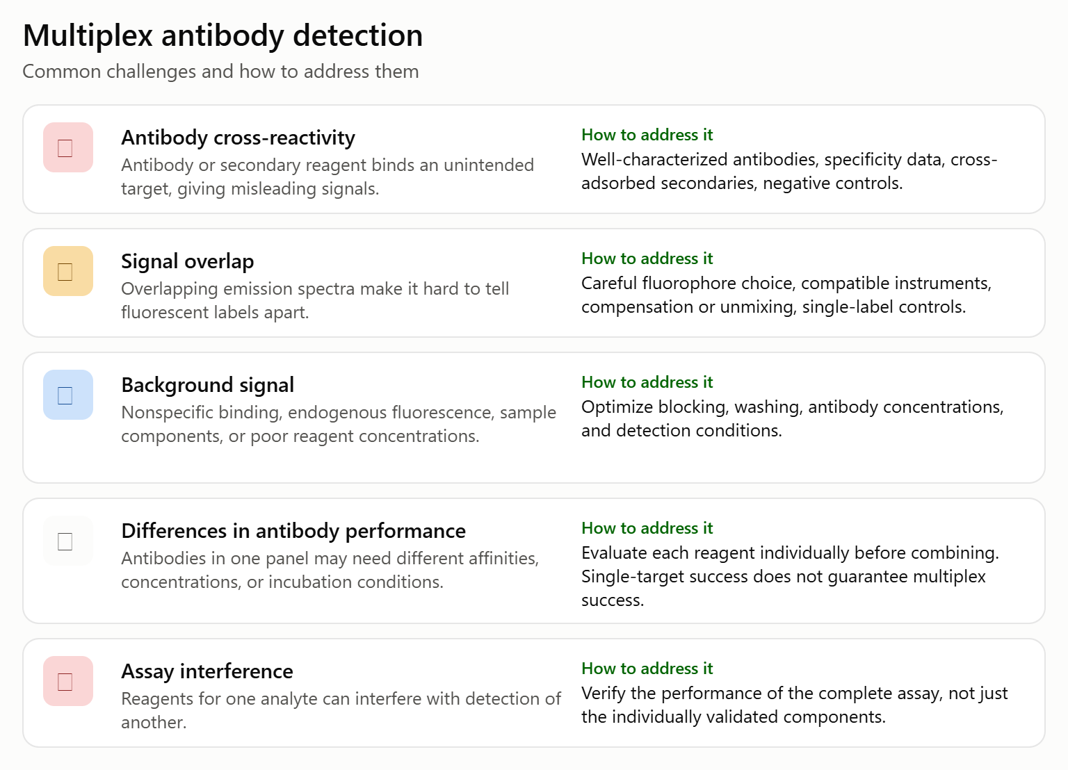
Multiplex antibody detection provides an effective approach for studying multiple biological targets within a shared experimental framework. By combining carefully selected antibodies with compatible detection technologies, researchers can investigate protein expression, cellular characteristics, biomarkers, and complex biological responses.
Reliable multiplex experiments depend on antibody specificity, host species compatibility, appropriate detection labels, signal separation, and rigorous controls. Understanding these factors helps laboratories design more effective assays and obtain results that can be interpreted with greater confidence.
As multiplex immunofluorescence, flow cytometry, and multiplex immunoassays continue to develop, antibody selection and validation remain central to high-quality biomedical research.